Microhematocrit · Measurement reliability
A microhematocrit centrifuge can finish its timed run and produce clearly separated blood layers without guaranteeing an accurate packed cell volume (PCV) result. Red cell packing, trapped plasma, capillary preparation, rotor-specific RCF and reader alignment must be evaluated separately.
Reliable PCV measurement requires more than successful centrifuge operation. The laboratory must establish that the sample was suitable, the packed red cell column met the method's requirements, and the final reading satisfied the applicable quality criteria.
THE CENTRAL DISTINCTION Visible separation is not sufficient packing. Sufficient packing is not proof of accuracy. Repeatable readings are not necessarily free from systematic bias.
01 · SAMPLE Pre-analytical integrity Anticoagulant, mixing, fill, seal and vessel compatibility.
02 · SPIN Packing conditions Rotor-specific RCF, time, loading and sample behaviour.
03 · READ Measurement quality Layer boundaries, reader alignment and method verification.
Clear Blood Layers Do Not Prove the PCV Is Correct
After a sealed capillary has been centrifuged, the sample commonly shows a packed red cell column near the sealed outer end, a narrow buffy coat containing mainly leukocytes and platelets, and plasma above. The spun packed cell volume is read as the proportion of the packed red cell column relative to the total blood column.
PCV (%) = (packed red cell column height ÷ total blood column height) × 100
The total blood column is measured from the bottom of the packed cells to the top of the plasma, excluding sealant. The buffy coat is not included in the packed red cell height. A 20 mm red cell column in a 50 mm total blood column gives a reading of 40%.
However, the calculation cannot tell whether the cells were adequately packed or whether the reader identified the correct boundaries. These must be considered separately.
| Observation | What It Establishes | What It Does Not Establish |
| Distinct cell and plasma layers | The sample has undergone visible separation. | That red cell packing is sufficient for the method. |
| A plateau after additional spinning | Further spinning produces no material change under the evaluated conditions. | That the result has no bias relative to an appropriate reference. |
| Close duplicate readings | Repeatability may be acceptable for that measurement set. | That both readings are free from a shared systematic error. |
Trapped Plasma: A Bias That Longer Spinning May Not Eliminate
As erythrocytes settle and pack, small spaces between cells can retain plasma. The observed packed red cell column is therefore not necessarily a perfectly pure volume of erythrocytes. Trapped plasma can make a spun PCV higher than the true cell volume fraction, and the extent of trapping may depend on sample characteristics, including abnormal red cell morphology.
Incomplete packing is different: under the selected conditions, the red cell column has not yet reached the method's required packing state, so additional spinning may still reduce its height. Residual trapped plasma may remain even when appropriate packing has been achieved.
Practical interpretation: If extending the spin changes PCV materially, review packing conditions. If the result has stabilised, do not assume every remaining difference from another method is a centrifugation-time problem. Trapped plasma and method-specific bias may still be relevant.
Anticoagulant and Capillary Selection Are Part of the Measurement
The pre-analytical process begins before the centrifuge starts. Capillary type must be matched to the collection method and the anticoagulation status of the sample.
DIRECT CAPILLARY COLLECTION
Anticoagulated capillary collection
When the method requires anticoagulation at direct collection, an appropriate heparinised microhematocrit capillary may be used. Select the specified tube rather than choosing by diameter alone.
PRE-ANTICOAGULATED BLOOD
Blood already collected into anticoagulant
EDTA-treated whole blood is commonly transferred into a plain capillary when this matches the validated method. The tube and anticoagulant combination should not be changed without checking the method.
Underfilling the primary collection tube can alter blood-to-anticoagulant ratio; liquid anticoagulants may also introduce dilution effects. Poor mixing, small clots, unsuitable storage or inconsistent capillary filling can contribute to unreliable results. A repeat spin cannot correct every error introduced before centrifugation.
Why the Same RPM Can Produce Different Centrifugal Force
RPM describes rotational speed, not the centrifugal force at a particular position in the rotor. RCF depends on rotational speed and radius:
RCF (×g) = 1.118 × 10 −5 × radius (cm) × RPM 2
The radius must be defined for the relevant rotor and measurement position. To illustrate, at 10,000 rpm, a theoretical radius of 7 cm corresponds to approximately 7,826 ×g, whereas 9 cm corresponds to approximately 10,062 ×g—about 28.6% greater force at the larger radius.
Calculation example, not an operating protocol: These radii are hypothetical and do not describe the GlanLab YT12M rotor. Neither result is a universal recommendation for microhematocrit testing.
When transferring a method between instruments or changing rotors, confirm the specified RCF, actual rotor geometry, compatible capillary tubes, run timing and the required performance checks. Copying the RPM setting alone does not reproduce a method's centrifugal conditions.
How Long Is Long Enough? What a 2025 Study Actually Demonstrated
A 2025 study in the Journal of Veterinary Diagnostic Investigation examined 188 blood samples from donkeys, dogs, sheep and cattle. The investigators compared results from shorter centrifugation times with results after a 15-minute reference spin, using approximately 14,972 ×g in the studied setup.
| Sample Group | Number of Samples | Shortest Studied Time Giving Agreement with the 15-minute Comparator |
| Donkeys | 44 | 3 minutes |
| Dogs | 43 | 3 minutes |
| Sheep | 61 | 10 minutes |
| Cattle | 40 | 5 minutes |
| Total | 188 | Specific to the study conditions |
The study assessed agreement with its 15-minute comparator, not absolute measurement truth. The sample population was also limited: all cattle and most donkey PCV values were within reference intervals, and polycythaemic animals were not included. The authors identified a need for further validation.
The useful lesson is not a species-specific timing prescription. It is that the packing behaviour of different blood samples may differ even under the same centrifugal conditions. Laboratories should establish and verify their own suitable method rather than assume that one timing rule applies to all samples or instruments.
Reader Alignment Can Change the Result After a Perfectly Executed Spin
The packed cell column must be read against the correct total column. In a standard reading approach, align the bottom of the red cell column with zero, the top of the plasma with the full-scale mark, and read the top of the packed red cells while excluding the buffy coat. Follow the exact reader's instructions for positioning and parallax control.
BOUNDARY ERROR
Including the buffy coat
Leukocytes and platelets in the buffy coat are not part of the packed red cell height. Counting the layer as red cells can raise the observed PCV.
DENOMINATOR ERROR
Counting the sealant
Sealant is not part of the original blood column. Incorrect alignment of the sealed end or plasma meniscus changes the apparent proportion.
POSITION ERROR
Misaligning the reader
Reader scale, tube position and viewing angle matter, particularly where the buffy coat is narrow or the meniscus is difficult to locate.
REPEATABILITY ERROR
Changing the reading procedure
Operator technique and timing should follow a consistent validated procedure. Inter-operator differences require evaluation.
For example, including an extra 0.5 mm as red cells in a 50 mm total blood column increases the apparent result by one percentage point (0.5 ÷ 50 × 100). This hypothetical example illustrates geometric reading bias; it is not a measured GlanLab performance value or a typical buffy-coat thickness.
Duplicate PCV Readings: Precision Is Not the Same as Accuracy
Duplicate capillaries can help reveal variability introduced by filling, sealing, centrifugation and reading. Whether duplicate results are acceptable should be judged against the laboratory's applicable method and quality criteria.
Two closely matching readings do not prove accuracy if both capillaries were exposed to the same incorrect RCF, the same sample problem or the same reader alignment error. Conversely, an unexpectedly large difference between duplicate readings calls for an investigation before a result is accepted.
Method verification may need to assess precision, bias against an appropriate comparison procedure, and performance across the sample range relevant to the intended use. A plateau with longer spinning, close duplicate readings and agreement with a validated reference address different questions.
Why Spun PCV and Automated Haematocrit May Differ
Spun PCV uses the height of the packed red cell column. Many automated haematology analysers derive haematocrit from red blood cell count and mean cell volume. The methods do not share identical error mechanisms.
Sample condition, red cell morphology, trapped plasma, analyser measurement effects and pre-analytical handling can influence their agreement. A difference between spun PCV and automated HCT is therefore not, by itself, evidence of centrifuge failure. Investigate the difference under the laboratory's method-comparison and quality-control procedures.
Where Should a Laboratory Investigate an Unexpected PCV Result?
| Observation | Check First | Avoid Assuming |
| PCV decreases after additional spinning | Whether initial packing was sufficient; actual RCF and time | That time is the only source of bias |
| Duplicate capillaries disagree | Mixing, filling, sealing, tube condition and reader technique | That the centrifuge motor is faulty |
| PCV appears systematically high | Trapped plasma, incomplete packing, buffy coat and method comparison | That longer spinning will remove all bias |
| Capillary breaks or leaks | Tube/rotor compatibility, seal and balanced loading | That the affected setup is safe for the next run |
| Spun PCV differs from automated HCT | Different method principles and sample-related effects | That either instrument must be defective |
| Results shift after rotor replacement | Radius, rotor-specific RCF, capillary position and transfer verification | That unchanged RPM means unchanged conditions |
These are investigation priorities, not automatic diagnoses. Abnormal results used in clinical decision-making require handling according to the laboratory's reporting and quality procedures.
What the GlanLab YT12M Specifications Mean for PCV Workflows
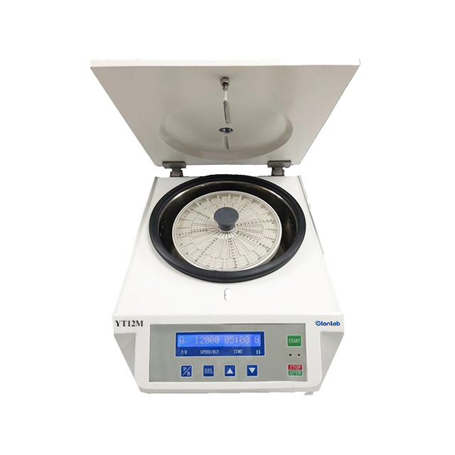
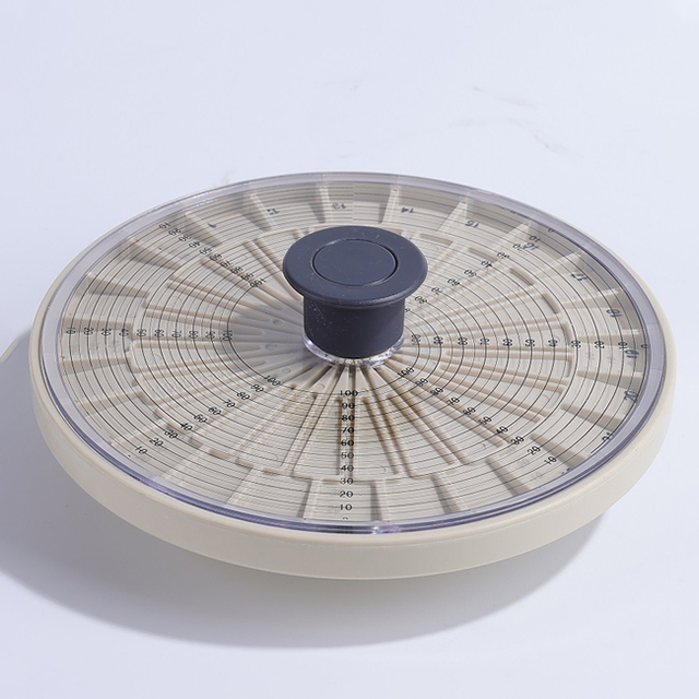
The GlanLab YT12M hematocrit centrifuge uses a dedicated 24-position capillary rotor with a haematocrit reader integrated into its rotor lid. Its published technical specifications define the machine's capability, not a guaranteed PCV accuracy for every sample or method.
| Published YT12M Specification | Value | What It Tells the Laboratory |
| Maximum speed | 12,000 rpm | Upper rotational capability, not the default PCV setting. |
| Maximum RCF | 15,800 ×g | Published maximum centrifugal force for the specified rotor configuration. |
| Capillary capacity | 24 positions | Maximum loading positions; balancing and tube compatibility still apply. |
| Speed accuracy | ±20 rpm | Manufacturer-published speed-control specification; not PCV result accuracy. |
| Timer | 0–99 min 59 s | Supports defined run timing. |
| Acceleration/deceleration profiles | 10 | Provides selectable motion control; use settings validated for the method. |
| Reading arrangement | Rotor-lid reader | Supports PCV reading within the matched accessory configuration. |
Specifications are not clinical performance claims. The YT12M's maximum speed, maximum RCF and speed accuracy do not independently demonstrate a particular PCV bias, diagnostic sensitivity or result precision. Those depend on the actual sample, capillary and validated measurement process.
When Consumables or Rotors Change, Reassess the Method
Capillary length, internal diameter, anticoagulant treatment, sealing material, rotor radius and reader geometry can all matter. Changing one component does not necessarily require full method redevelopment, but it does require determining which compatibility and performance checks are affected.
A physically fitting capillary is not automatically approved for high-speed use. Likewise, a replacement rotor with a different radius can change RCF at the same RPM. Confirm the approved tube/rotor combination and the appropriate verification before routine use.
For equipment purchasing questions—capillary dimensions, rotor capacity, instrument configuration and reader arrangements—see the Microhematocrit Centrifuge Buying Guide .
The Four Controls Behind a Trustworthy PCV Measurement
1 · Sample preparation Correct anticoagulation, mixing, filling, sealing and sample condition.
2 · Centrifugation Approved capillary/rotor, actual RCF, run time and balanced loading.
3 · Reading Correct blood-column alignment, buffy-coat exclusion and reader technique.
4 · Quality verification Suitable precision, bias and method-performance checks for the intended use.
A finished spin proves only that the cycle ended. It does not, by itself, establish that the cells were adequately packed, the blood column was read correctly or the final result met the laboratory's acceptance criteria. Reliable microhematocrit testing depends on treating the centrifuge, capillaries, reader, samples and validated procedure as one measurement system.
Technical References
Clinical and Laboratory Standards Institute (CLSI). Procedure for Determining Packed Cell Volume by the Microhematocrit Method, H07-A3, third edition, 2000. Archived technical reference.
World Health Organization / International Council for Standardization in Haematology. Recommended Method for the Determination of Packed Cell Volume by Centrifugation, 2000.
Journal of Veterinary Diagnostic Investigation, 2025, 37(6):873–881. Estimation of Minimum Centrifugation Time of Microhematocrit Tubes to Obtain Accurate Results of Packed Cell Volume and Total Solids in Donkeys, Dogs, Sheep, and Cows.
GlanLab. YT12M Hematocrit Centrifuge — Published Technical Specifications.
Use the applicable validated method, laboratory SOP and vessel/rotor instructions for actual operating conditions.
Check Your Microhematocrit Equipment Configuration
Share the capillary type, sample workflow, required RCF and reading requirements to discuss the GlanLab YT12M and its matched accessories.
View YT12M